Introduction
Ligament injuries are among the most common musculoskeletal conditions evaluated in orthopedic and sports medicine clinics. Whether sustained during competitive athletics, an unexpected fall, a motor vehicle collision, or a sudden awkward twisting motion, damage to these dense connective tissues can compromise joint function and cause sudden pain and swelling.To assist patients and families in researching clinical options, platforms such as BESTORTHOHOSPITALS provide structured directories of orthopedic surgical centers, sports medicine departments, and rehabilitation facilities. While online directories serve as helpful educational tools, they do not independently certify clinical outcomes or replace an in-person clinical examination by a licensed orthopedic specialist.
What Are Ligaments?
Ligaments are dense, fibrous bands of collagenous connective tissue that connect one bone to another across a synovial joint. Composed primarily of closely packed, parallel bundles of Type I collagen fibers interspersed with specialized cells called fibroblasts (ligamentocytes), ligaments serve essential mechanical and sensory functions.
                      ┌─────────────────────────────────────────┐
                      │      Primary Functions of Ligaments     │
                      └────────────────────┬────────────────────┘
                                           │
         ┌─────────────────────────────────┴─────────────────────────────────┐
         ▼                                                                   ▼
┌────────────────────────────────┐                  ┌────────────────────────────────┐
│      Mechanical Stability      │                  │   Proprioceptive Feedback      │
│ Prevents excessive, abnormal   │                  │ Houses mechanoreceptors that   │
│ joint translations, rotations, │                  │ send positional data to the    │
│ and unnatural hyper-extensions │                  │ brain to coordinate balance    │
└────────────────────────────────┘                  └────────────────────────────────┘
Examples of Major Ligament Complexes:
- Knee Ligaments:
- Anterior Cruciate Ligament (ACL): Resists anterior translation of the tibia and rotational forces.
- Posterior Cruciate Ligament (PCL): Resists posterior displacement of the tibia relative to the femur.
- Medial Collateral Ligament (MCL): Stabilizes the inner knee against valgus (inward) stress.
- Lateral Collateral Ligament (LCL): Stabilizes the outer knee against varus (outward) stress.
- Ankle Ligaments:
- Anterior Talofibular Ligament (ATFL) & Calcaneofibular Ligament (CFL): Stabilize the lateral ankle against inversion (rolling inward) injuries.
- Deltoid Ligament Complex: Provides medial ankle stability against eversion stress.
- Upper Extremity Ligaments:
- Ulnar Collateral Ligament (UCL) of the Elbow: Resists valgus stress, commonly stressed in throwing athletes.
- Glenohumeral Ligaments: Provide static stabilization to the ball-and-socket joint of the shoulder.
Grading the Severity of a Ligament Injury
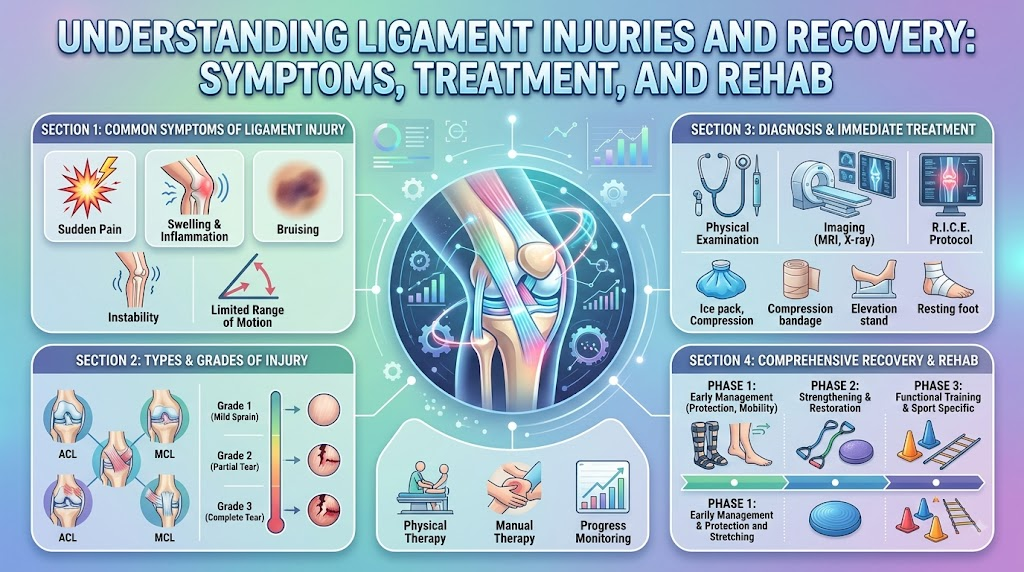
In orthopedic medicine, ligament injuries are clinically classified as sprains and graded on a three-tier scale based on structural disruption and mechanical joint stability:
┌─────────────────────────────────────────────────────────────────────────────┐
│                       Clinical Grading of Ligament Sprains                  │
├──────────────────────┬────────────────────────┬─────────────────────────────┤
│ Severity Grade       │ Pathological Disruption│ Clinical & Joint Findings   │
├──────────────────────┼────────────────────────┼─────────────────────────────┤
│ Grade 1 (Mild)       │ Microscopic stretching │ Localized tenderness; mild  │
│                      │ of collagen fibers;    │ swelling; no mechanical laxity│
│                      │ no gross structural tear│ or joint instability        │
├──────────────────────┼────────────────────────┼─────────────────────────────┤
│ Grade 2 (Moderate)   │ Incomplete, partial    │ Moderate pain, swelling, and│
│                      │ tearing of the ligament│ bruising; mild to moderate  │
│                      │ fibers                 │ joint laxity with solid stop│
├──────────────────────┼────────────────────────┼─────────────────────────────┤
│ Grade 3 (Severe)     │ Complete rupture or    │ Significant swelling; gross │
│                      │ avulsion from the bone │ mechanical joint instability│
│                      │ attachment             │ ("giving way"); no end point│
└────────────────┴────────────────────────┴─────────────────────────────┘
A precise physical examination and diagnostic imaging are necessary to distinguish between grades, as clinical treatment pathways diverge based on joint laxity.
Common Causes and Mechanisms of Injury
Ligaments are engineered to withstand substantial tensile forces, but they can fail when external loads exceed the structural limits of their collagen fibers.
                      ┌───────────────────────────────────────┐
                      │    Primary Mechanisms of Injury       │
                      └──────────────────┬────────────────────┘
                                         │
     ┌──────────────────┬────────────────┼────────────────┬──────────────────┐
     ▼                  ▼                ▼                ▼                  ▼
┌──────────┐     ┌─────────────┐  ┌─────────────┐  ┌─────────────┐    ┌─────────────┐
│ Non-     │     │ Direct      │  │ Awkward     │  │ Sudden      │    │ High-Energy │
│ Contact  │     │ Blunt Force │  │ Landing     │  │ Deceleration│    │ Traumatic   │
│ Pivoting │     │ Impact      │  │ Mechanics   │  │ & Cutting   │    │ Accidents   │
└──────────┘     └─────────────┘  └─────────────┘  └─────────────┘    └─────────────┘
- Non-Contact Deceleration and Pivoting: Rapidly changing direction while running or landing with a dynamic valgus collapse (knees buckling inward) is the classic mechanism for ACL injuries.
- Direct Blunt Impact: A direct blow to the lateral aspect of the knee (such as a tackle in football) forces the joint inward, stressing or tearing the MCL.
- Inversion of the Ankle: Stepping on an uneven surface or landing on an opponent’s foot forces the ankle into plantarflexion and inversion, stressing the lateral ankle ligament complex (ATFL/CFL).
- Hyperextension or Dashboard Impact: Direct force applied to the anterior tibia when the knee is bent (e.g., a car dashboard impact) forces the shin backward, injuring the PCL.
- Repetitive Valgus Overload: High-velocity overhead throwing motions generate micro-trauma across the elbow UCL over time.
Recognizing Common Symptoms
The symptoms of a ligament injury vary depending on the joint affected and the severity of the tear:
- Acute Pain: Immediate, sharp pain localized to the joint line or site of ligament attachment at the moment of injury.
- Rapid Onset Swelling (Hemarthrosis): Swelling that develops within 1 to 4 hours of injury often indicates bleeding inside the joint capsule, frequently seen with intra-articular tears like the ACL.
- Audible or Palpable “Pop”: Many patients report hearing or feeling a distinct snap or pop at the time of injury.
- Joint Instability and “Giving Way”: A sensation that the joint is loose, buckled, or unable to support body weight during pivoting or normal walking.
- Ecchymosis (Bruising): Visible purple-blue discoloration that tracks down the limb over 24 to 48 hours as internal blood settles.
- Loss of Range of Motion: Difficulty bending, extending, or fully rotating the affected joint due to swelling, pain, or protective muscle spasms.
When to Consult an Orthopedic Professional
While mild muscle aches often resolve with rest, suspected ligament injuries warrant a formal clinical assessment to rule out severe tears or associated joint damage:
- Inability to bear weight on the injured leg or take four steps immediately following the trauma.
- Sensation of the knee, ankle, or shoulder shifting, sliding out of place, or giving way.
- Rapid, tense swelling developing within hours of the incident.
- Joint pain or mobility restrictions that fail to improve after 5 to 7 days of home care.
- Persistent joint stiffness or recurrent catching, locking, or clicking sensations during movement.
- High-demand athletes planning a return to pivoting, cutting, or jumping sports.
Emergency Warning Signs: Ruling Out Severe Complications
Certain clinical signs indicate potential fractures, joint dislocations, or neurovascular compromise that require immediate hospital emergency room evaluation:
🚨 SEEK EMERGENCY MEDICAL CARE IMMEDIATELY IF YOU OBSERVE: 🚨

• Obvious anatomical deformity, visible bone protrusion, or gross joint misalignment.
• Inability to feel the foot, hand, or toes (numbness, tingling, or complete loss of sensation).
• Pale, blue-tinted, or cold skin distal to the injured joint with absent or faint pulses.
• Sudden, extreme, uncontrollable pain disproportionate to the injury (suspected compartment syndrome).
• Inability to move the digits (toes or fingers) distal to the injured site.
• Open lacerations or puncture wounds communicating with a swollen, injured joint (open joint injury).
How Orthopedic Specialists Diagnose Ligament Injuries
A comprehensive orthopedic evaluation follows a systematic diagnostic pathway:
┌─────────────────────────────────────────────────────────────────────────────┐
│                       Orthopedic Diagnostic Pathway                         │
├──────────────────────────────────────┬──────────────────────────────────────┤
│ 1. Clinical History                  │ • Determining the exact mechanism,   │
│                                      │   weight-bearing status, and "pop"   │
├──────────────────────────────────────┼──────────────────────────────────────┤
│ 2. Physical Examination              │ • Palpation of joint lines;          │
│                                      │   provocative joint stability tests  │
├──────────────────────────────────────┼──────────────────────────────────────┤
│ 3. Plain Radiography (X-Rays)        │ • Evaluates for avulsion fractures,  │
│                                      │   dislocations, and bone alignment   │
├──────────────────────────────────────┼──────────────────────────────────────┤
│ 4. Magnetic Resonance Imaging (MRI)  │ • High-resolution soft-tissue imaging│
│                                      │   of ligaments, cartilage, & meniscus│
└──────────────────────────────────────┴──────────────────────────────────────┘
Specialized Physical Stress Tests
Orthopedic clinicians perform specialized, non-invasive manual stability tests to evaluate ligament integrity:
- Knee: Lachman Test and Pivot-Shift Test (ACL), Posterior Drawer Test (PCL), Valgus/Varus Stress Tests (MCL/LCL).
- Ankle: Anterior Drawer Test and Talar Tilt Test (ATFL/CFL).
- Elbow: Valgus Stress Test and Moving Valgus Stress Test (UCL).
Role of Diagnostic Imaging
While plain X-rays do not directly display soft-tissue ligament fibers, they are essential to rule out fractures or bone avulsions (where the ligament pulls away a small fragment of bone). Magnetic Resonance Imaging (MRI) remains the gold standard for visualizing soft tissues, assessing tear severity, and checking for associated meniscus, labral, or chondral (cartilage) damage.
Initial Care Principles (PEACE & LOVE Framework)
In modern sports medicine, the traditional RICE protocol has evolved into the more comprehensive PEACE & LOVE framework, emphasizing active, protected recovery over prolonged, passive immobilization:
┌─────────────────────────────────────────────────────────────────────────────┐
│                         Immediate Care: P.E.A.C.E.                          │
├─────────────────────────────────────────────────────────────────────────────┤
│ [P] Protect   ──► Unload and restrict movement for the first 1 to 3 days.   │
│ [E] Elevate   ──► Elevate the injured limb higher than the heart for fluid. │
│ [A] Avoid     ──► Avoid anti-inflammatory medications and excessive ice     │
│                   that may impair natural tissue healing cascades.          │
│ [C] Compress  ──► Use elastic bandages to minimize intra-articular edema.   │
│ [E] Educate   ──► Learn active recovery strategies from a healthcare team.  │
├─────────────────────────────────────────────────────────────────────────────┤
│                         Subsequent Care: L.O.V.E.                           │
├─────────────────────────────────────────────────────────────────────────────┤
│ [L] Load      ──► Introduce active movement and mechanical loading early.   │
│ [O] Optimism  ──► Maintain a positive, realistic outlook to aid recovery.   │
│ [V] Vascular  ──► Choose pain-free cardiovascular exercise to boost flow.   │
│ [E] Exercise  ──► Perform progressive strength, mobility, and balance drills.│
└─────────────────────────────────────────────────────────────────────────────┘
Note: Initial care guidelines provide temporary comfort while awaiting clinical evaluation; they do not replace formal orthopedic assessment.
Non-Surgical (Conservative) Management Pathways
Many ligament injuries heal successfully without surgical intervention. The potential for non-operative healing depends on the ligament’s intrinsic vascularity. For example, the Medial Collateral Ligament (MCL) and lateral ankle ligaments have robust blood supplies and surrounding vascular soft tissues, allowing most Grade 1, Grade 2, and even select isolated Grade 3 tears to heal conservatively.
Core Components of Conservative Management:
- Immobilization & Bracing: Using hinged knee braces, walking boots, or lace-up ankle supports to protect healing collagen fibers from abnormal lateral or rotational strain while allowing safe, linear movement.
- Protected Weight-Bearing: Using crutches temporarily to reduce compressive axial joint loads during the acute inflammatory phase.
- Early Guided Physical Therapy: Initiating safe range-of-motion drills early to prevent arthrofibrosis (joint stiffness) and muscle inhibition.
- Neuromuscular Re-Education: Restoring proprioception and stabilizing the joint through muscle co-contraction (strengthening the hamstrings, quadriceps, calves, or peroneal muscles to compensate for ligament laxity).
When Is Surgery Considered?
Surgery is not required for every ligament tear, but it becomes an important consideration under specific clinical circumstances:
                      ┌───────────────────────────────────────┐
                      │    Indications for Surgical Review    │
                      └──────────────────┬────────────────────┘
                                         │
     ┌──────────────────┬────────────────┼────────────────┬──────────────────┐
     ▼                  ▼                ▼                ▼                  ▼
┌──────────┐     ┌─────────────┐  ┌─────────────┐  ┌─────────────┐    ┌─────────────┐
│ Gross,   │     │ High-Demand │  │ Multi-      │  │ Associated  │    │ Failed      │
│ Recurrent│     │ Pivoting    │  │ Ligament    │  │ Meniscal /  │    │ Conservative│
│ Joint    │     │ Athletic    │  │ Knee Injuries│ │ Cartilage    │    │ Rehab Trial │
│ Laxity   │     │ Goals       │  │ (Dislocation)│ │ Damage      │    │ (Instability)│
└──────────┘     └─────────────┘  └─────────────┘  └─────────────┘    └─────────────┘
- Intra-Articular Ligament Ruptures with Poor Vascularity: The ACL is bathed in synovial fluid and lacks a vascular sheath; complete ruptures rarely heal spontaneously. Active individuals experiencing functional instability often choose surgical stabilization.
- Multi-Ligament Joint Injuries: High-energy traumas involving simultaneous tears (e.g., combined ACL, MCL, and PCL injuries) require surgical repair or reconstruction to prevent severe chronic joint laxity.
- Associated Repairable Structural Damage: Tears occurring alongside bucket-handle meniscus tears or chondral defects that require arthroscopic repair.
- High-Level Athletic or Occupational Demands: Individuals whose professions or competitive sports require high-speed pivoting, cutting, or heavy physical labor.
Understanding Ligament Reconstruction vs. Repair
When surgery is indicated, orthopedic surgeons utilize specialized techniques based on the tissue quality and injury location:
| Surgical Approach | Primary Mechanism | Typical Clinical Applications |
| Ligament Repair | The torn ligament ends are sutured directly back together or re-anchored to the bone using suture anchors. | Acute bony avulsion fractures, proximal MCL tears, or select pediatric/adolescent tears with high tissue quality. |
| Ligament Reconstruction | The ruptured ligament tissue is excised and replaced with a tissue graft routed through precise anatomical bone tunnels. | Complete ACL tears, chronic PCL ruptures, complex lateral ankle reconstructions, or revised ligament surgeries. |
┌─────────────────────────────────────────────────────────────────────────────┐
│                         Types of Reconstruction Grafts                      │
├───────────────────────────────────┬─────────────────────────────────────────┤
│ Autografts (Patient's Own Tissue) │ • Bone-Patellar Tendon-Bone (BPTB)      │
│                                   │ • Hamstring Tendon (Semitendinosus)     │
│                                   │ • Quadriceps Tendon                     │
├───────────────────────────────────┼─────────────────────────────────────────┤
│ Allografts (Donor Tissue)         │ • Donor Achilles, Tibialis, or Patellar │
│                                   │   tendons; useful for multi-ligament    │
│                                   │   reconstructions or revision surgeries │
└───────────────────────────────────┴─────────────────────────────────────────┘
Physiotherapy and Rehabilitation: The Four Phases
Rehabilitation is the central pillar of ligament recovery, whether managed conservatively or post-surgically. Modern rehabilitation follows a criteria-based progression (advancing based on functional milestones rather than strict calendar days).
┌─────────────────────────────────────────────────────────────────────────────┐
│                      Phases of Ligament Rehabilitation                      │
├──────────────────────┬────────────────────────┬─────────────────────────────┤
│ Phase 1: Protection  │ Phase 2: Neuromuscular │ Phase 3 & 4: Functional &   │
│ & Mobility           │ Strength               │ Sports Return               │
├──────────────────────┼────────────────────────┼─────────────────────────────┤
│ • Reduce swelling &  │ • Restore full active  │ • Dynamic single-leg balance│
│   manage pain        │   range of motion      │ • Plyometrics & agility     │
│ • Restore full knee  │ • Progressive closed-  │ • Sport-specific drills &   │
│   extension/mobility │   chain strength drills│   limb symmetry index >90%  │
│ • Quadriceps wake-up │ • Core & pelvic kinetic│ • Psychological readiness   │
│   (prevent arthrogenic│   chain integration   │   assessment (ACL-RSI)      │
│   muscle inhibition) │                        │                             │
└──────────────────────┴────────────────────────┴─────────────────────────────┘
Phase 1: Joint Protection, Swelling Control, and Early Mobility
- Goals: Resolve swelling, restore normal joint range of motion (especially full, passive extension in the knee), and reactivate inhibited muscles through isometric contractions and straight-leg raises.
Phase 2: Progressive Strength and Neuromuscular Control
- Goals: Build baseline muscular strength across the entire kinetic chain using closed-kinetic-chain exercises (squats, lunges, leg presses, step-downs) and balance boards to re-educate mechanoreceptors.
Phase 3: Dynamic Stability, Plyometrics, and Agility
- Goals: Introduce impact-absorption drills, multi-directional cutting, hopping, and decelerating mechanics while ensuring proper knee-over-toe alignment without dynamic valgus collapse.
Phase 4: Sport- or Work-Specific Conditioning and Return to Activity
- Goals: Replicate sport-specific movements, build high-speed cardiovascular conditioning, complete objective functional testing batteries, and assess psychological confidence.
What Influences the Recovery Timeline?
Ligament healing is an ongoing biological process. Because collagen remodeling takes time, patients should understand the key biological and individual factors that influence the overall timeline:
┌─────────────────────────────────────────────────────────────────────────────┐
│                     Factors Governing the Healing Timeline                  │
├───────────────────────────────────┬─────────────────────────────────────────┤
│ 1. Ligament Vascular Supply       │ Extra-articular tissues (MCL/ATFL) heal │
│                                   │ faster than intra-articular (ACL/PCL).  │
├───────────────────────────────────┼─────────────────────────────────────────┤
│ 2. Biological Graft Remodeling    │ Reconstructed grafts undergo necrosis,  │
│    ("Ligamentization")            │ revascularization, and cellular         │
│                                   │ remodeling over 9 to 18 months.         │
├───────────────────────────────────┼─────────────────────────────────────────┤
│ 3. Associated Structural Trauma   │ Multi-tissue injuries (e.g., meniscus   │
│                                   │ repair) require protected early phases. │
├───────────────────────────────────┼─────────────────────────────────────────┤
│ 4. Adherence to Rehabilitation    │ Consistency with daily exercises and    │
│                                   │ movement quality directly guides pace.  │
└───────────────────────────────────┴─────────────────────────────────────────┘
While a mild Grade 1 ankle sprain may resolve within 2 to 4 weeks, a surgically reconstructed ACL typically requires 9 to 12 months of structured rehabilitation before safe clearance for pivoting sports can be granted.
Returning to Work: Practical Considerations
The timeline for returning to occupational duties depends on the physical demands of your specific job role:
- Sedentary / Desk-Based Roles: Many patients return within 1 to 3 weeks, provided pain is controlled, the limb can be elevated comfortably, and commute logistics are manageable.
- Standing or Light-Walking Jobs: Roles involving light retail or teaching typically require 4 to 8 weeks, depending on joint stability, swelling control, and tolerance for standing.
- Heavy Manual Labor & Construction: Careers involving ladder climbing, heavy lifting, squatting, and uneven ground work often require 4 to 9 months, alongside formal physical therapy work-conditioning programs.
Returning to Sports: Why Objective Testing Matters
Returning to sport should never be based solely on how many months have passed since the injury or surgery. Feeling pain-free during daily walking does not mean the joint is ready for high-velocity cutting, jumping, or contact.
Objective Return-to-Sport Criteria Battery:
- Limb Symmetry Index (LSI) > 90–95%: Strength testing (isokinetic dynamometry or hand-held dynamometry) demonstrating that the injured limb has at least 90–95% of the strength of the uninjured limb.
- Functional Single-Leg Hop Testing: Passing a battery of single-leg hop tests (single hop for distance, triple hop, crossover hop, timed 6-meter hop) with an LSI > 90%.
- Movement Quality Assessment: Video analysis confirming proper trunk, hip, and knee alignment during drop vertical jumps, without inward knee collapse.
- Psychological Readiness: Validated scoring on tools like the ACL-Return to Sport after Injury (ACL-RSI) scale, confirming the athlete has confidence and is free of debilitating fear of reinjury.
Common Mistakes to Avoid During Recovery
- Returning to Pivoting Sports Too Early: Returning to competition before completing full neuromuscular rehabilitation and meeting objective strength benchmarks, significantly increasing reinjury risks.
- Stopping Rehabilitation When Pain Resolves: Assuming that the absence of pain equals full ligament healing, dynamic stability, and muscle recovery.
- Neglecting Kinetic Chain Strength: Focusing only on the injured joint while ignoring hip abductors, gluteals, and core stabilizers that control lower-limb alignment.
- Immobilizing the Joint for Too Long: Keeping a joint completely immobilized without medical direction, leading to arthrofibrosis, muscle atrophy, and joint stiffness.
- Skipping Warm-Ups and Pre-Activity Conditioning: Failing to perform dynamic neuromuscular warm-ups (such as the FIFA 11+ program) before athletic activities.
- Comparing Timelines with Others: Assuming your recovery will match a friend’s or professional athlete’s pace, overlooking differences in surgical techniques, tissue quality, and individual healing.
Systemic and Lifestyle Factors Affecting Healing
Tissue repair is supported by general physiological and lifestyle health:
- Nutrition and Protein Intake: Adequate dietary protein (1.2 to 2.0 grams per kilogram of body weight during active rehab) supplies essential amino acids for collagen synthesis and muscle preservation.
- Hydration: Proper hydration maintains synovial fluid viscosity and supports cellular nutrient transport across recovering tissues.
- Sleep and Restorative Recovery: Growth hormone and tissue-repair cytokines are predominantly released during deep, non-REM sleep cycles; aiming for 7 to 9 hours of quality sleep supports healing.
- Tobacco and Nicotine Avoidance: Nicotine causes peripheral vasoconstriction, reduces tissue oxygen delivery, and impairs collagen synthesis, significantly delaying healing and increasing surgical non-union rates.
- Metabolic Health: Managing underlying systemic conditions like diabetes mellitus and maintaining blood glucose control supports microvascular perfusion and reduces infection risks.
How to Choose a Quality Orthopedic Hospital
When selecting a specialized hospital, medical center, or sports medicine institute for ligament care, look for facilities that provide comprehensive, multi-specialty services:
                      ┌───────────────────────────────────────┐
                      │   Leading Orthopedic Center Pillars   │
                      └──────────────────┬────────────────────┘
                                         │
     ┌──────────────────┬────────────────┼────────────────┬──────────────────┐
     ▼                  ▼                ▼                ▼                  ▼
┌──────────┐     ┌─────────────┐  ┌─────────────┐  ┌─────────────┐    ┌─────────────┐
│ Board-   │     │ High-Res    │  │ Minimally   │  │ Integrated  │    │ Functional  │
│ Certified│     │ 3T MRI &    │  │ Invasive    │  │ Dedicated   │    │ Testing &   │
│ Orthopedic│    │ Digital     │  │ Arthroscopy │  │ Sports      │    │ Transparent │
│ Surgeons │     │ Diagnostics │  │ Theaters    │  │ Rehab Units │    │ Care Plans  │
└──────────┘     └─────────────┘  └─────────────┘  └─────────────┘    └─────────────┘
- Board-Certified Specialists: Orthopedic surgeons with dedicated subspecialty fellowship training in Sports Medicine, Arthroscopy, and Joint Reconstruction.
- Advanced Diagnostic Infrastructure: On-site high-resolution 1.5T or 3T MRI systems and digital radiography for accurate soft-tissue visualization.
- Modern Arthroscopic Surgical Suites: Fully equipped operative rooms with advanced fluid-management systems, high-definition visualization, and modern anatomical fixation devices.
- Integrated Physical Therapy & Rehabilitation: On-site rehabilitation departments staffed by certified sports physiotherapists who utilize criteria-based protocols and objective functional testing equipment.
- Multidisciplinary Collaborative Care: Close coordination between orthopedic surgeons, sports medicine physicians, radiologists, physical therapists, and athletic trainers.
Hospital Evaluation & Research Matrix
Use this structured matrix when evaluating prospective orthopedic hospitals, sports medicine centers, or specialized surgical clinics:
| Assessment Factor | Hospital A | Hospital B | Hospital C |
| Orthopedic Surgeon Qualifications | Board-Certified / Sports Fellowship | Board-Certified / Sports Fellowship | Board-Certified / Sports Fellowship |
| On-Site 3T / 1.5T Diagnostic MRI | Available | Available | Available |
| Minimally Invasive Arthroscopic Surgery | Dedicated suites | Dedicated suites | Dedicated suites |
| Integrated Physical Therapy Department | On-site with sports gym | On-site with sports gym | On-site with sports gym |
| Objective Return-to-Sport Testing | Force plates / Hop tests / Iso | Force plates / Hop tests / Iso | Force plates / Hop tests / Iso |
| Cartilage & Meniscus Preservation Focus | Available | Available | Available |
| Emergency Orthopedic Trauma Access | 24/7 coverage | 24/7 coverage | 24/7 coverage |
| Transparent Package & Billing Estimates | Itemized written quote | Itemized written quote | Itemized written quote |
| Accreditation (e.g., NABH, JCI, AAAHC) | Documented | Documented | Documented |
Myths vs. Facts About Ligament Injuries
| Myth | Fact |
| Myth: Every complete ligament tear requires surgical repair or reconstruction. | Fact: Many complete tears (such as isolated MCL or ankle ligament tears) heal well with conservative bracing and targeted physical therapy. |
| Myth: If you can walk on an injured joint, it is definitely not torn. | Fact: Many individuals can walk in straight lines with a torn ACL or ankle ligament, but experience instability during pivoting or rotational forces. |
| Myth: Once pain disappears, the ligament is completely healed and ready for sports. | Fact: Pain resolves long before collagen fibers fully reorganize and dynamic neuromuscular strength, balance, and stability are restored. |
| Myth: An MRI is always mandatory immediately following every minor joint sprain. | Fact: Orthopedic specialists use clinical history and physical exams to determine when an MRI is necessary, avoiding unnecessary scans for mild sprains. |
| Myth: Wearing a brace permanently eliminates the risk of future ligament tears. | Fact: Braces provide external support, but they cannot replace neuromuscular strength, proper biomechanics, and active joint stability. |
| Myth: Reconstructed ligaments are ready for full sports after 6 months. | Fact: Contemporary evidence shows that biological graft remodeling and neuromuscular control often require 9 to 12 months to lower reinjury rates. |
How BESTORTHOHOSPITALS.COM Can Support Your Research
Navigating musculoskeletal injuries and understanding clinical treatment options is an essential first step toward full recovery. BESTORTHOHOSPITALS.COM offers organized educational resources and directories to assist patients:
- Orthopedic Hospital & Clinic Discovery: Search directories of specialized orthopedic surgical centers, academic sports medicine institutes, and joint reconstruction departments.
- Explore Clinical Procedures: Access evidence-based overviews of arthroscopic ACL reconstruction, collateral ligament repair, ankle stabilization, and cartilage restoration.
- Destination Research: Review hospital infrastructure, surgical technology, and rehabilitation capabilities across various medical hubs.
- Consultation Preparation: Use structured checklists and question templates to prepare for detailed discussions with your orthopedic team.
Platform Note: BESTORTHOHOSPITALS.COM is an educational healthcare directory designed for research and educational purposes. The platform does not provide individualized medical diagnoses, guarantee surgical outcomes, or replace professional clinical evaluations. All medical decisions should be made in direct consultation with a qualified orthopedic specialist.
Frequently Asked Questions (FAQs)
1.What is a ligament injury?
A ligament injury occurs when strong bands of fibrous connective tissue connecting bones across a joint are stretched, partially torn, or completely ruptured due to sudden twisting, trauma, or abnormal mechanical force.
2.What are the symptoms of a torn ligament?
Common symptoms include sudden joint pain, rapid swelling, audible or palpable popping sensations at the moment of injury, bruising, restricted movement, and a feeling that the joint is unstable or giving way under body weight.
3.What are the different grades of ligament injuries?
Ligament injuries are graded as Grade 1 (mild microscopic stretching with no joint laxity), Grade 2 (moderate partial fiber tearing with slight laxity), and Grade 3 (severe, complete ligament rupture or detachment with significant joint instability).
4.Can a ligament injury heal without surgery?
Yes. Many ligament injuries—particularly extra-articular tears like MCL sprains and lateral ankle sprains—have a rich blood supply and heal successfully with bracing, activity modification, and structured physical therapy.
5.How long does ligament recovery take?
Recovery varies widely based on injury severity and location; mild Grade 1 sprains often heal within 2 to 4 weeks, while complex tears or surgically reconstructed ligaments (like the ACL) typically require 6 to 12 months of structured rehabilitation.
6.When should I see an orthopedic specialist?
You should see an orthopedic specialist if you experience severe joint pain, rapid swelling, inability to bear weight, visible deformity, persistent joint instability, or if symptoms fail to improve after several days of home care.
7.Do ligament injuries always require an MRI?
Not always. A thorough physical exam and X-rays are often sufficient for initial assessment, though an MRI is recommended when a complete tear, surgical candidate, or associated meniscus or cartilage injury is suspected.
8.What is the role of physiotherapy in ligament recovery?
Physiotherapy is essential for restoring joint mobility, reducing swelling, rebuilding muscle strength, re-educating neuromuscular proprioception, and preparing the patient for safe return to daily activities and sports.
9.When can I return to sports after a ligament injury?
Return to sports should be criteria-based, requiring full range of motion, normal joint stability, limb strength symmetry above 90%, passing dynamic hop tests, and demonstrating psychological readiness rather than relying solely on elapsed time.
10.Can I exercise with a ligament injury?
Yes, but exercise must be carefully structured and non-provocative. Early in recovery, pain-free upper-body workouts, stationary cycling, or guided isometric strengthening may be performed under clinical supervision while protecting the injured joint.
11.What factors affect ligament recovery?
Recovery is influenced by the specific ligament injured, severity grade, associated cartilage or meniscus damage, patient age, vascular supply, adherence to physical therapy, nutrition, sleep quality, and complete avoidance of smoking.
12.What are the warning signs of a serious ligament injury?
Warning signs requiring urgent medical evaluation include visible joint deformity, severe numbness or loss of sensation in the foot or hand, cold or pale extremities, absent distal pulses, and uncontrolled pain.
13.How do I choose a hospital for ligament treatment?
Look for an accredited hospital staffed by fellowship-trained sports medicine orthopedic surgeons with access to high-resolution MRI diagnostics, advanced arthroscopic theaters, and an on-site, criteria-based physical therapy department.
14.What questions should I ask an orthopedic surgeon?
Ask about the specific grade of your tear, whether non-surgical management is appropriate for your athletic goals, graft options if surgery is needed, expected rehabilitation timelines, and objective criteria required for sports clearance.
15.How can BESTORTHOHOSPITALS.COM help me research orthopedic hospitals?
BESTORTHOHOSPITALS.COM provides organized directories of orthopedic hospitals, procedural overviews, and consultation preparation tools to help you research facilities and prepare for clinical visits.
Conclusion
Understanding ligament injuries and recovery is the foundation for restoring joint stability, functional mobility, and long-term musculoskeletal health. Because ligaments vary in their anatomical location, blood supply, and mechanical demands, successful healing requires an individualized clinical approach rather than a generic timeline.